Tissuegnostics Info Letter – March 2021

Tissuegnostics Precision that inspires

Colon Cancer Biomarker

Tissue cytometry as tool for prognostic biomarker detection in cancer

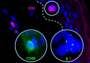
A study conducted by a group from the University of Western Australia, published in the journal Cancer Science, explores spatial relationships and the prognostic impact of PD-L1+ dendritic cells and CD8+ T cells on survival of colon cancer patients. The contextual image analysis solution StrataQuest was used for the biomarker assessment.
» get the pdf article

Graft Versus Host Disease

Webinar: In-situ Immunophenotyping in graft-versus host disease

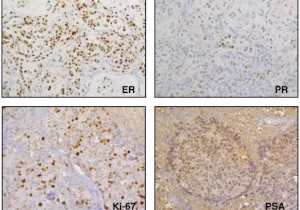
Dr. Johanna Strobl, PhD, from the Medical University of Vienna, Austria, talks about her recently published study addressing host-derived skin-resident memory T cells as potential players in human graft versus host disease. The acquisitions/identification of skin-resident memory T cells in-situ was performed with tissue cytometry solutions.
» watch the webinar

Disseminated Cancer Cells

Assessment of inducers of disseminated cancer cell progression

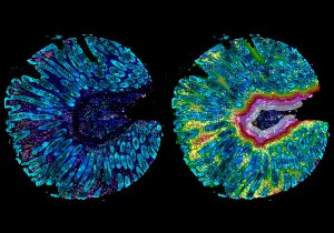
The group of Prof. Klein (University of Regensburg, Germany) published a paper in Nature Communications investigating initial steps of metastasis formation in breast cancer by performing a comprehensive analysis of disseminated cancer cells and microenvironmental signals in xenografts. TissueFAXS i PLUS and HistoQuest were used to determine expression of various markers.
» get the pdf article

Image Cytometry Solutions

Enhance your research with AI-assisted tissue cytometry

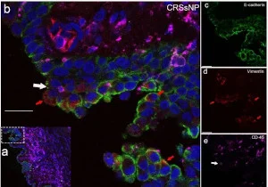
Use TissueGnostics automated analysis solutions to choose from a multitude of analysis options: high-content (immuno)phenotyping by multiplexing/spectral unmixing, spatial context analysis, unlimited tissue classification by machine learning, molecular single-cell profiling using RNA ISH, imaging mass cytometry, pathogen quantification etc.
» watch a short video on tissue cytometry

Chronic Rhinosinusitis

Comprehensive cellular characterization of chronic rhinosinusitis

The group of Giotakis et al (Medical University of Innsbruck, Austria) performed an in-depth analysis of the cellular composition within human chronic rhinosinusitis. Various cell populations, including epithelial-, connective tissue-, and immune cells were characterized in-situ using TissueFAXS and the contextual image analysis solution StrataQuest.

» get the pdf article

Precision that inspires. Explore the fundamental unit of life

Heliosonix

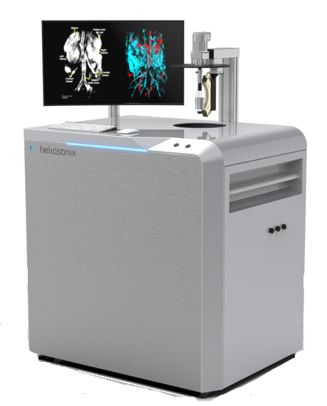
Heliosonix AREA
Heliosonix AREA

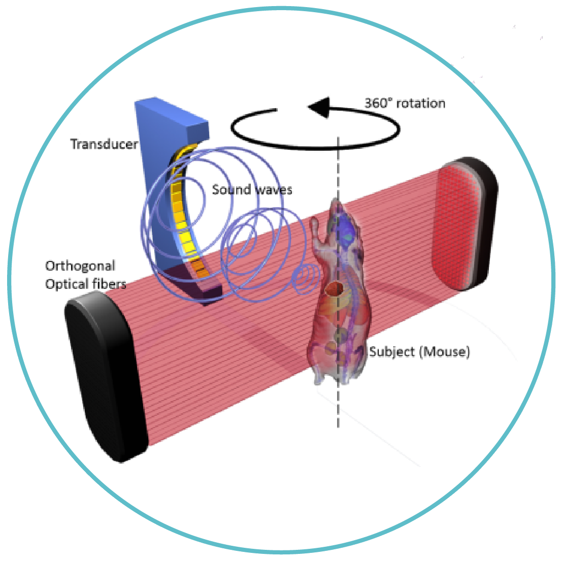
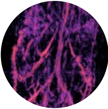
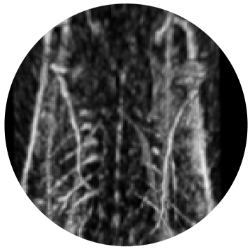
When a short-pulsed laser energy penetrates the tissue, it heats and expands, generating thermo-acoustic (ultrasound) waves, which can be reconstructed to show the distribution of optical absorption inside the tissue.

Keeps mice alive – under anaesthesia
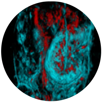
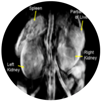
Obtains live 3D images with high resolution of inner structures,
including organ, vasculature and skeleton systems
Captures bones, soft tissue organs and blood flow
Delivers results within in 3-4 min
Allows to quantitatively track the effect of pharma
treatment over time in the same animal

AREA does not require regulatory approval
Buyers: universities, CROs, biopharma
Numerous prototype delivered and installed
Commercial sales launched in 2020 and 2 systems
sold and installed in China

TissueFAXS Fluo

TissueFAX FLUO

TissueFAXS Fluo
UPRIGHT FLUORESCENCE SLIDE SCANNER

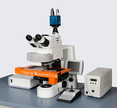
TissueFAXS Fluo is an open concept, widefield, upright, fluorescence slide scanner designed for slides, cytospins, smears and tissue microarrays. The fluorescence slide scanning system is equipped with TissueQuest or StrataQuest Fluo image analysis software of which there are no equals.

UNIQUE FEATURES

  • Fluorescence slide scanning & analysis
  • 8 capacity slide scanning
  • Slide label ID recognition
  • Quantitative image analysis option
  • TMA & FISH scanning & analysis
  • Automatic tissue detection
  • Extended focus, stitching & illumination correction
  • Image and metadata storage, management and archiving
  • Completely upgradable to higher capacity and Confocal slide scanning
  • Over 1,500 publications reference TissueGNOSTICS fluorescence slide scanners
TissueFAX FLUO

For additional information, call Meyer Instruments, Inc. at 1 (281) 579-0342 to speak with a representative.

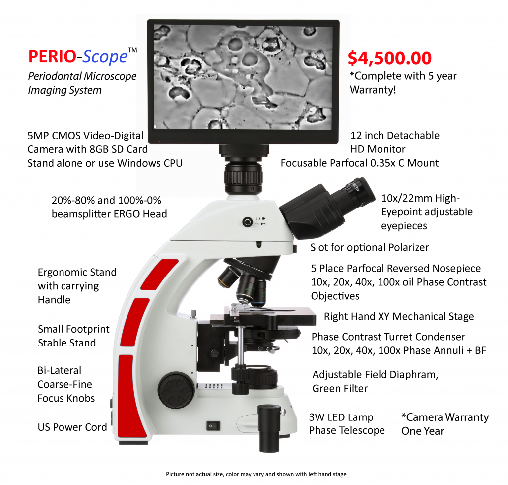
Announcing PERIO-Scope

PERIO-Scope microscope imaging system for PERIODONTISTS

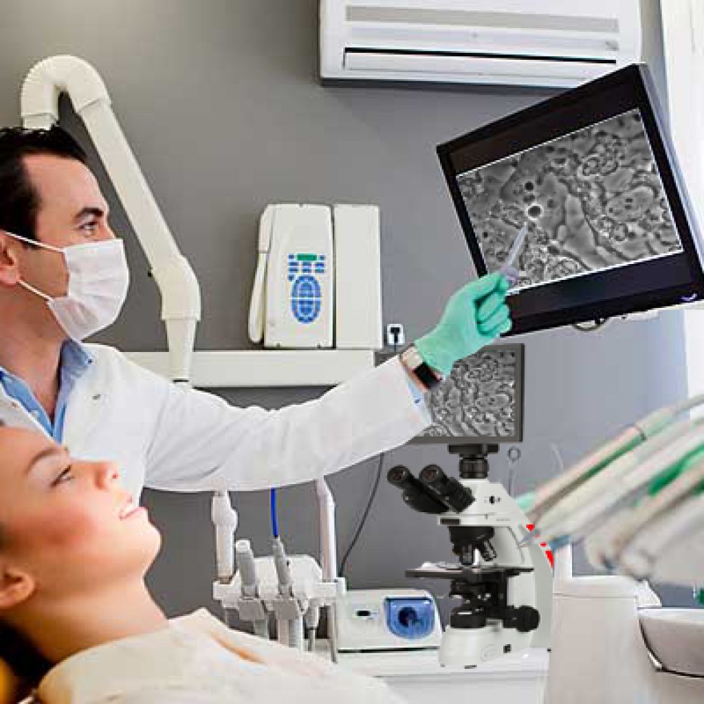
Our new product is designed specifically for periodontists to help prevent further gum disease by showing their patients how to achieve a healthy mouth with ‘LIVE’ video imaging. “If a picture is worth a thousand words, video is worth a million, seeing is believing”

  • New way to monitor and help eliminate periodontal gum disease.
  • Easy to use and amazing to watch your patient’s reaction!

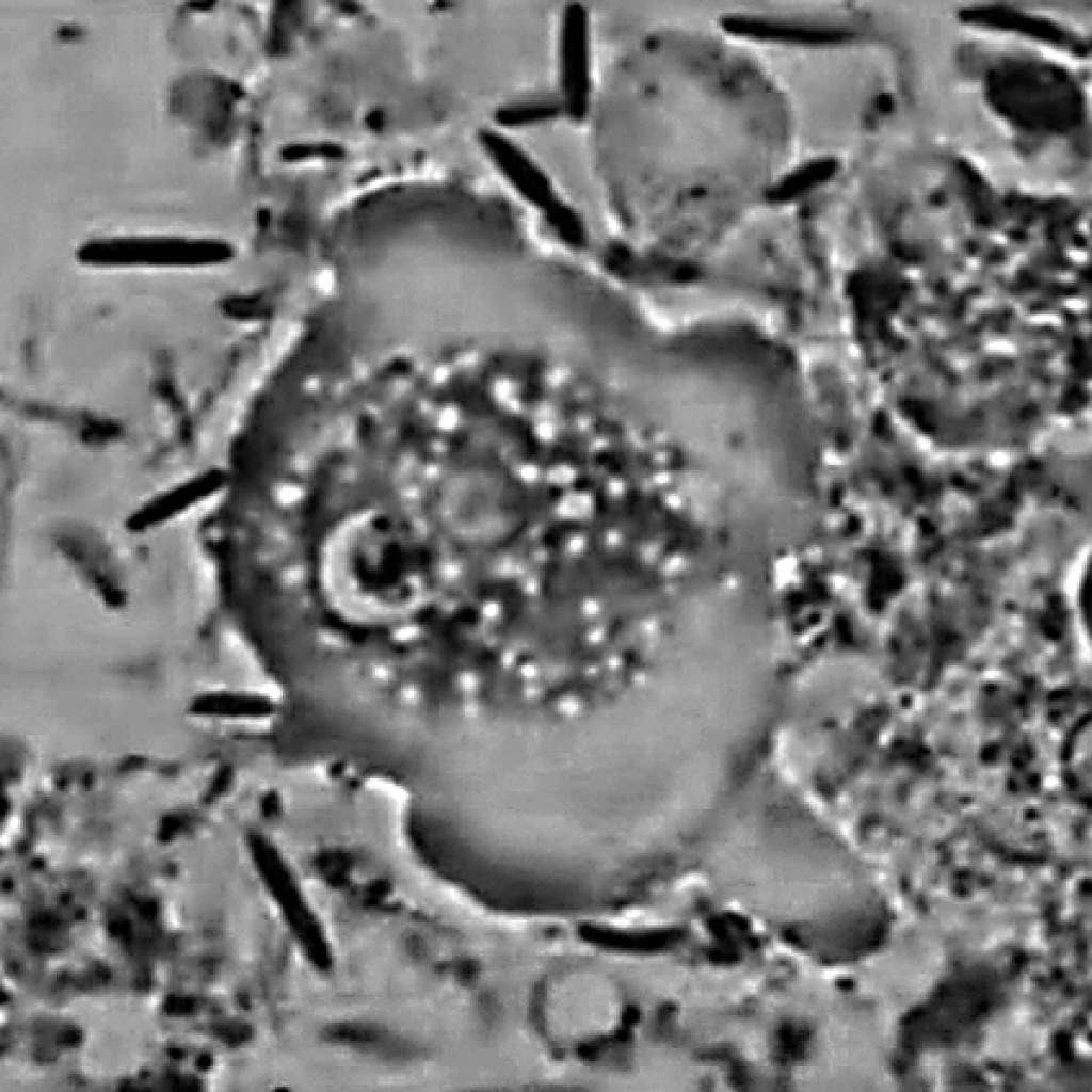
The PERIOScope is designed to allow patients to see these living organisms in their own mouths.

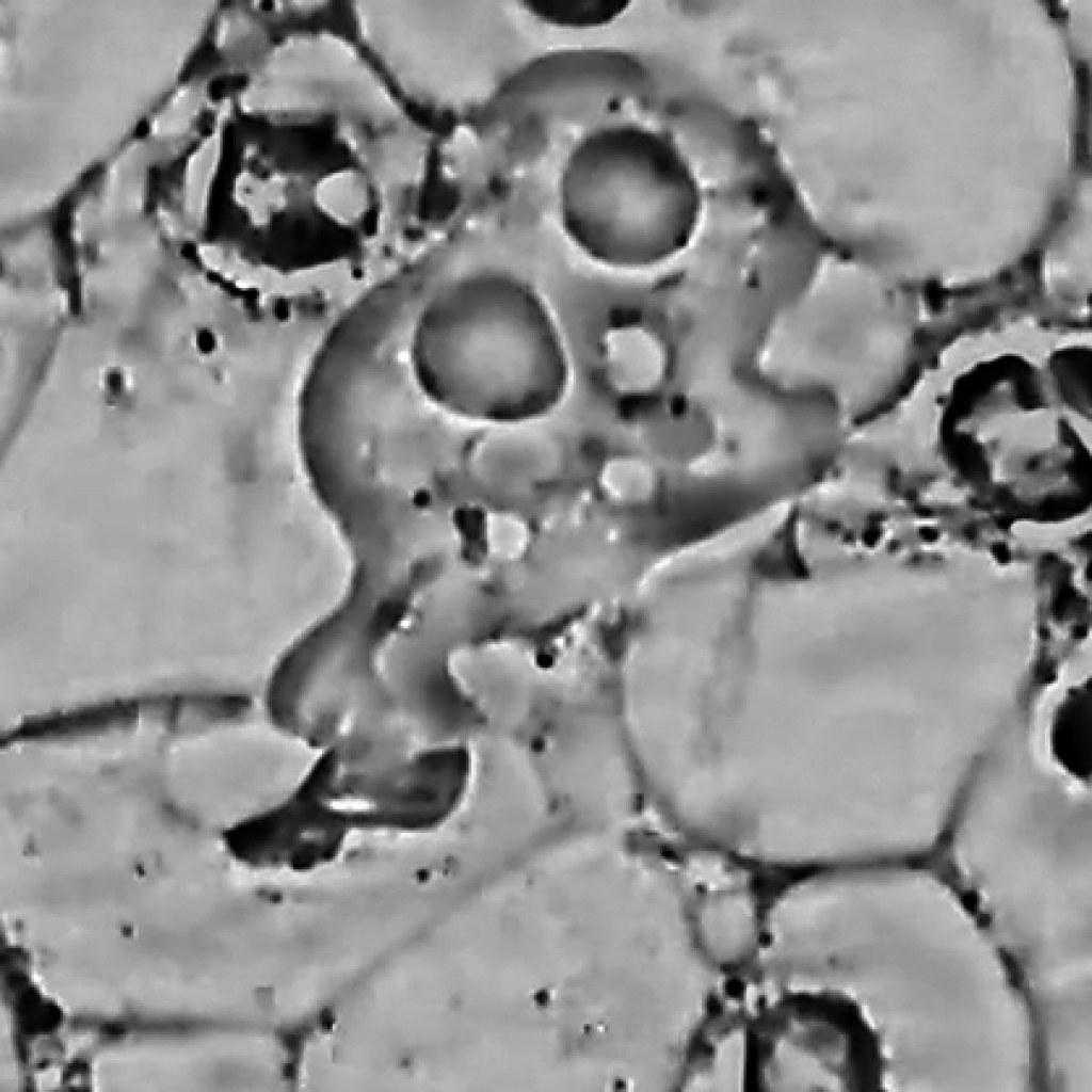
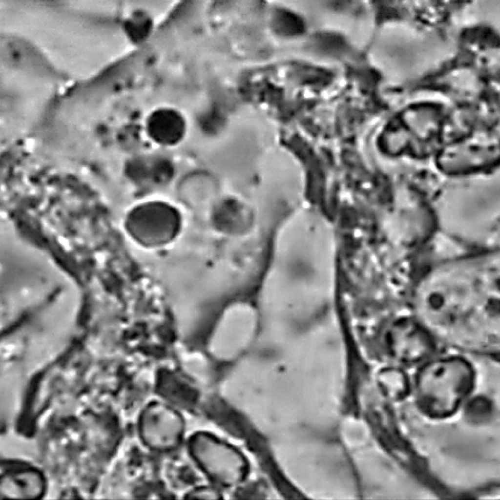
Periodontal Disease treatment can be visually explained to your patients with the help of a dedicated microscope and video camera capable of recording live video and still image capture. When images of epithelial cells, bacteria, red and white blood cells, trichomonas and even amoebas are shown to patients they can see for themselves the severity of gum disease.

Our PERIOScope phase contrast microscope imaging system is a simple, effective way for periodontists to show the ill effects of gum disease. Using “LIVE’ video feed, patients can experience for themselves the long-term effects of poor dental hygiene.

Using our specialized imaging tools, periodontists can visualize, record and track the progress of treatment. Operation is easy and fast. When used in conjunction with large HD monitors the images almost jump off the screen for patients and health providers benefit and understanding.

The PERIOScope comes complete and ready to use stand-alone with the included 12-inch HD monitor or with a Windows or Mac computer connected via USB2 cable. Beautiful live, phase contrast images of unstained saliva showcase all manner of material found in the patient’s mouth and gums.

Motic Launching FS-Live

Call Meyer Instruments for the Motic FS Live

Motic Launching FS-Live Telepathology System for Frozen Sections

Motic announces the FS-Live Telepathology System for frozen cases or fine-needle aspiration. This advanced, microscope-based whole slide imaging system provides high quality live images efficiently to pathologists from anywhere in the world, specifically for remote diagnosis and interpretation. The FS-Live eliminates the need to send pathologists and other health specialists to remote hospitals solely to read these cases. The system allows any pathologist on the network to view a case slide in a matter of seconds. Available with single or 6 slide capacity.

“Motic is relentless in perfecting the art of telepathology and digital pathology,” said Alex Lobozar, Motic General Manager, Sales and Marketing for North America. “We are constantly exploring ways to improve our instruments to be increasingly user-friendly and accessible for all.”

Key Features

Continuous Autofocus – unique to ALL scanners on the market, SUPER time saver

Live Mode 2x, 4x, 10x, 20x, 30x, 40x – duplicates common magnifications

Moisture Resistant – can be used with wet slides

Simple Operation – no complicated instructions to learn

Keyboard, Mouse or Hot Key Controls – multiple ways to focus or change magnifications

Heat Map – keep track of where you have viewed and at what magnification

Acts as a traditional scanner as well – save slides in multiple formats SVS, TIFF, JPEG, JPEG 2000, MDS


HREM – New 3D Imaging

HREM 3D

Call Meyer Instruments at 281-463-0342 for more information about this amazing 3D Digital Imaging System.

High Resolution Episcopic Microscope (HREM)

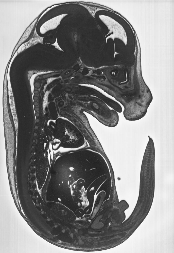
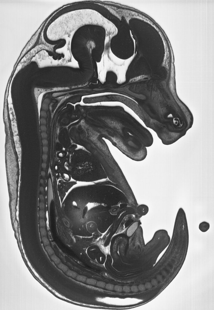
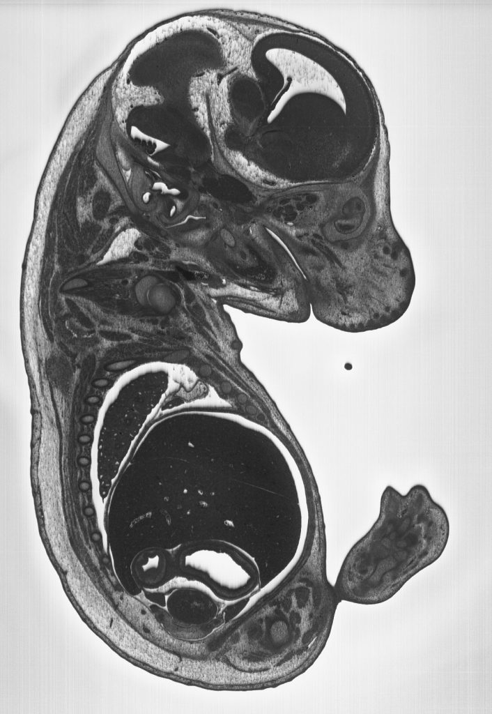
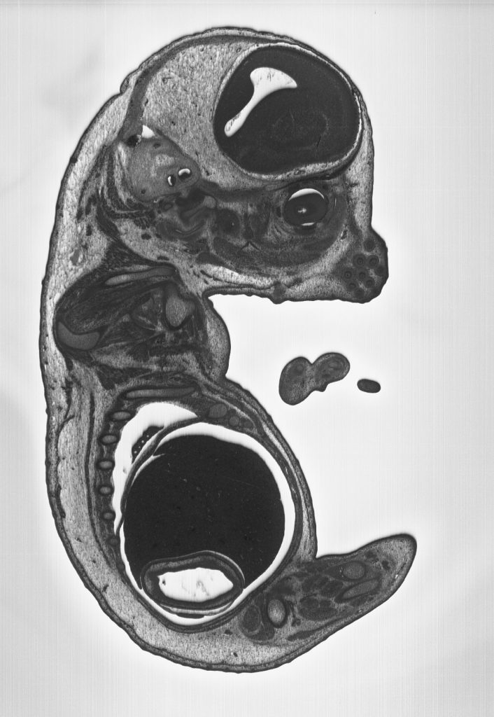
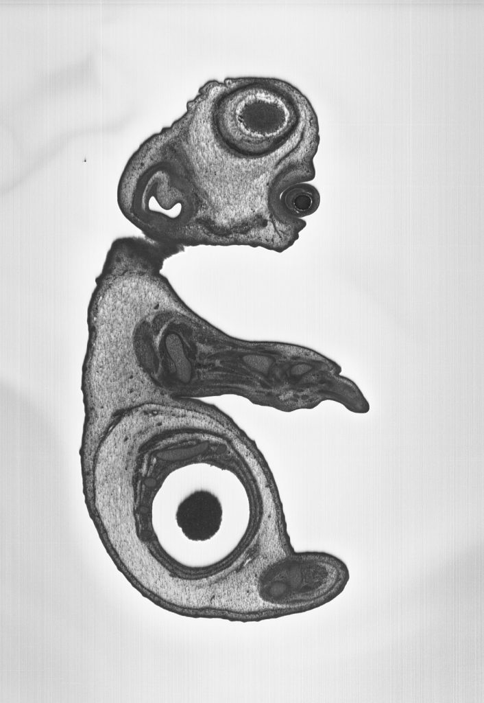
The optical High Resolution Episcopic Microscope (HREM) is a 3 dimensional image generation device for whole histological samples or embryo’s.

Consisting of a motorized microtome, fluorescence stereomicroscope and camera, the HREM cuts away a sample at 1-5 micron intervals and photographs the remaining faceplate, section by section. The result is hundreds or thousands or perfectly aligned images that are available for reassembling in 3D. Maximum sample size is 25mm in diameter (although they are experimenting with a new 40mm holder as well)

Microscopy and Image Analysis Publication

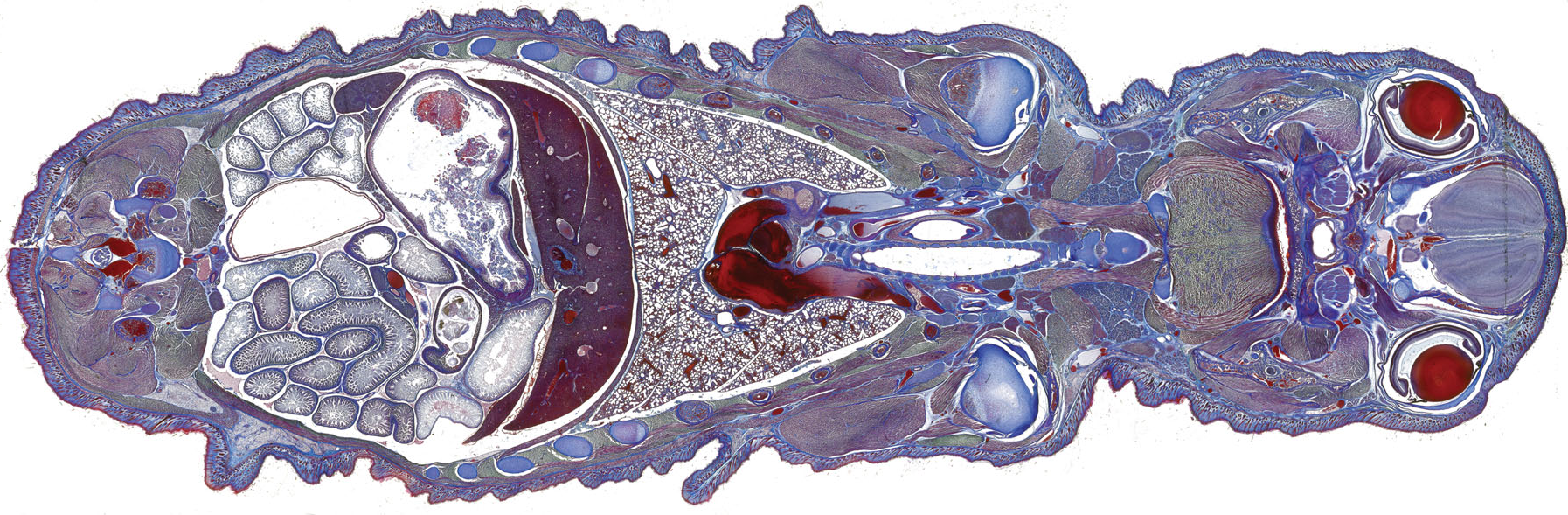
This publication is an overview of light microscopy, including objectives, light sources, filters, film, and color photography for fluorescence microscopy and fluorescence in situ hybridization (FISH). Features an image scanned with a Meyer Instruments, Inc., Pathscan Enabler 5, see more information below.

Microscopy Image Analysis

Download Article on Microscopy and Image Analysis

UNIT 4.4 Microscopy and Image Analysis- George McNamara1, Michael Difilippantonio2, Thomas Ried3, Frederick R. Bieber4 – Published Online: 11 JUL 2017

Pathscan Enabler 5

The PathScan Enabler 5 is the latest version of the patented PathScan series digital slide scanners. The original PathScan Enabler helped create the market for personal digital slide scanning over 20 years ago. With the new intuitive design, the PathScan Enabler 5 allows scanning of whole mounted histological samples attached to standard 1 X 3 inch cover slipped glass slides. The new optional GEOLOGICAL POLARIZING holder allows scanning of polished rock thin sections attached to 1 X 2 inch glass slides in both brightfield and polarized light!
The PathScan Enabler 5 solves low power, large field of view, and high resolution imaging problems. Scan area is 24.3 x 36.5 mm, resulting in stunning, incredible images up to 14,320 x 9,522 pixels! The PathScan Enabler 5 holds two (2) 1 X 3 inch glass slides and includes simple to use software for both Windows and Mac computers. The GEOLOGICAL POLARIZING holder accepts one (1) 1 x 2 inch glass slide. Image files are saved in TIFF or JPEG formats. The PathScan Enabler 5 is designed not to replace but complement a microscope and digital camera. The entire sample is scanned with an even, clear background and makes a perfect OVERVIEW image that can be viewed in a variety of software programs. Image scans are equivalent to 50X microscope resolution. Now in its 20th year of production, thousands are in use!

For more information See Meyer Instruments Website at https://meyerinst.com/pathscan-enabler-5/ on contact:

Meyer Instruments, Inc.

4202 Bear Lodge Court

Houston, TX 77084

281-579-0342

Meyer Sample Photo on GigaMacro

GIGAmacro’s Picture of the Month uses our own sample.

Take a look at this psychedelic image and guess what it is.

So what is it? It is a rock – a very thin piece of rock, but rock nonetheless. Andesite Thin Section, imaged at 10X with a Full Waveplate. We worked with Rob Meyer, our distributor in Houston, TX to develop the techniques to make this image. The thin section is lit from below by polarized flash, and the waveplate is inserted into a holder in the lens stack.

A common use of waveplates—particularly the sensitive-tint (full-wave) and quarter-wave plates—is in optical mineralogy. Adding waveplates between the polarizers of a petrographic microscope makes easier the optical identification of minerals in thin sections of rocks,[2] in particular by allowing deduction of the shape and orientation of the optical indicatrices within the visible crystal sections. This alignment can allow discrimination between minerals which otherwise appear very similar in plane polarized and cross polarized light. (source: wikipedia).

The original image is 1.94 gigapixels in size, 42,435 x 45,656 pixels at 58,521 pixels per inch. Made using 9,108 photos in 18 rows | 11 columns | 46 stacks.

As always, be sure to dig around by zooming and panning around in this image (or any others) in the GIGAmacro Viewer.

Learn more about this fascinating imagery and how it is made. Contact Meyer Instruments, Inc., (281) 579-0342 or visit their website at https://meyerinst.com.

Meyer Instruments, Inc. is an authorized dealer reseller of GIGAmacro products.