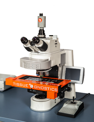
TissueFAXS Histo
UPRIGHT BRIGHTFIELD SLIDE SCANNER
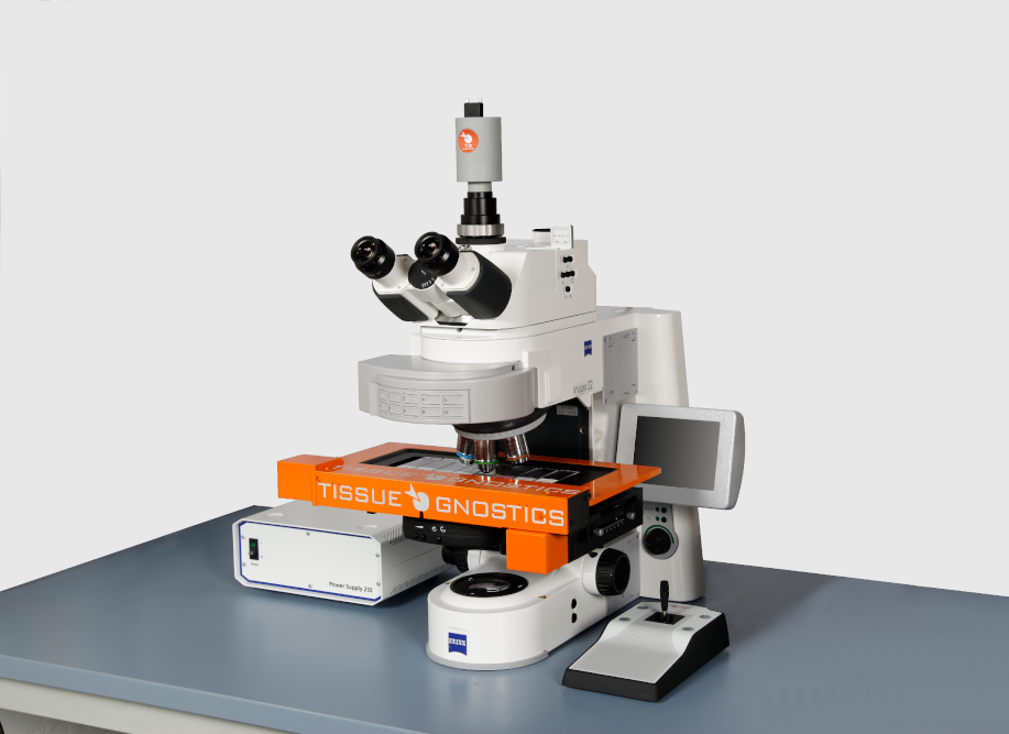
TissueFAXS Histo is an upright brightfield only system for the scanning and analysis of slides, cytospins, smears and Tissue Microarrays. The system is equipped with HistoQuest and/or StrataQuest Histo image analysis solftware.
 TissueFAXS Histo is also available in a scan only configuration.
 TissueFAXS Histo can be upgraded to the TissueFAXS PLUS configuration.
TissueFAXS Histo
UNIQUE FEATURES
- Brightfield scanning
- Automated tissue detection
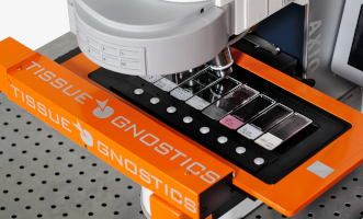
- 8 slides scanning
- Slide ID Scanner
- Quantitative image analysis option
- TMA & CISH scanning & analysis
- Extended focus, stitching & illumination
- Image and metadata storage, management and archiving
CONFIGURATION
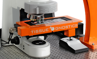
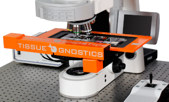
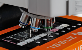
TissueFAXS Histo is TG upright brightfield system. The system scans and analyses samples on slides and slide-based sample containers in brightfield mode. It comes equipped with an 8-slide stage.
The objective turret allows for up to 7 objectives from all Zeiss objective classes with a M24 thread. TG continuously checks the market for cameras and other technical equipment to provide those solutions that deliver top performance and are cost-effective. Our current TissueFAXS Histo standard camera is a color 8 bit CMOS camera.
TissueFAXS Scanning and Management software integrates all the hardware components into an easy to use workflow with full, walk-away automation capabilities.
TissueFAXS Histo
UPGRADES
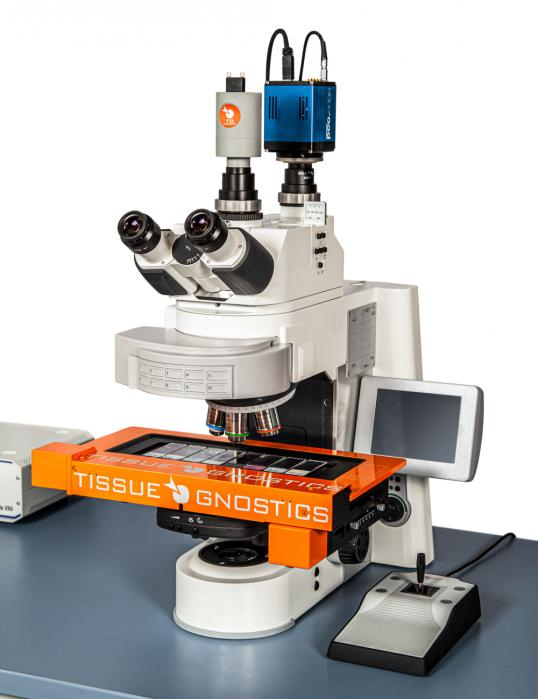
All imaging systems are modular and upgradable. Every system can be customized to offer the following capabilities: brightfield scanning, widefield fluorescence, confocal, and multispectral. To exemplify TissueFAXS Histo i can be ugraded to
– TissueFAXS PLUS (Image on the left)
– TissueFAXS Q+
– TissueFAXS SPECTRA
– TissueFAXS SL
PROPERTIES
Design
Modular (Microscope, Camera, Light-source, PC with 2 Monitors)
Microscopy mode
Brightfield
Compatible Slide Formats
All standard and over-sized slides
Slide Capacity
8
Objectives
Up to 7 Objectives (2.5-100x)
Camera
CMOS Camera (8/10-bit, 4.2 Megapixel, Color)
Light Source
VIS-LED
Tissue Microarray (TMA)
YES
Image Analysis Software
QUEST line (Algorithms for tissue diseases, Area measurment, scattergrams for cells and many more)
Supported Image Formats
TissueFAXS, OME-TIFF, TIFF, JPEG, BMP, PNG
Viewer
Integrated, stand-alone freeware

