StrataQuest APP analysis examples
In the following some examples for analyses done with StrataQuest APPs are shown.
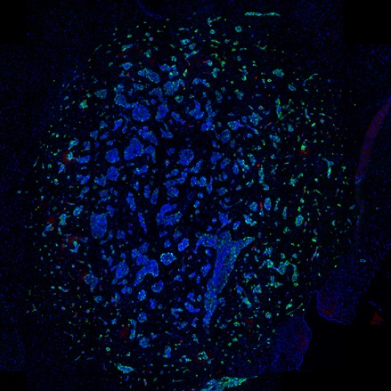
IF Immune Status in situ APP: Distance of tumor cells from blood vessels
The following example was analysed using a combination of the IF Immune Status in siu APP and the FL version of the IHC Angio APP. It was performed for the Johns Hopkins Hospital in New York. The aim was to evaluate the distance of tumor cells from blood vessels within the tumor cell clusters.
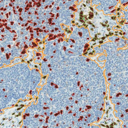
IHC Meta.Cells APP: Stroma and tumor detection on morphological stains
One of the main strengths of StrataQuest is the capability to segment morphological structures based on a morphological stain alone (i.e. without a specific staining of the structure to be detected). In this case, the task of segmentation into tumor and stroma was made more difficult due to many very thin areas of stroma.
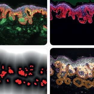
Analysis of cyclic stain and bleach samples
Multi-epitope ligand cartography (MELC) is a technology using samples subjected to cycles of fluorescent staining, imaging and photobleaching. Each cycle can use an antibody for a different protein. The result is a set of images of the distributions of many proteins for the same samples.
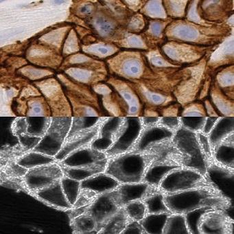
IHC Membrane APP: HER2/neu in Breast Cancer
Within this project the aim was to measure the prominent diagnostic parameter for breast cancer, the intensity of the HER2/neu membrane staining and the completeness of the closure of the respective membrane rings.
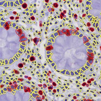
IHC2 APP: Ki-67 nuclear staining analysis
Ki-67 staining and evaluation is an integral part of cancer diagnosis. Despite it being routinely used in pathology there still are issues to its correct use. Automated tissue analysis can provide a valuable supplement to visual evaluation. The IHC2 APP is among the StrataQuest easiest to use APPs and requires very little preparation time.
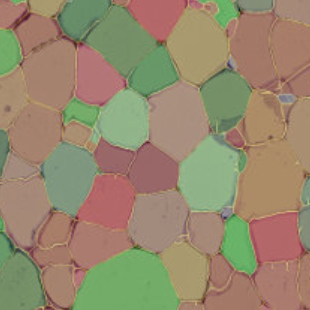
Adipocyte APP: Measurement of cell size
The Adipocyte APP was designed from the ground up to be run in routine by staff untrained in image analysis and image processing. The picture set is loaded into StrataQuest and the results are calculated. The algorithm automatically eliminates adipocytes on the borders from the analysis. Minor membrane tears, inevitable in working with adipose tissue sections, are automatically closed by the analysis algorithm. Larger sections of missing membrane and cell membranes collapsed into the lumina can be drawn in, respectively out using simple manual painting tools.
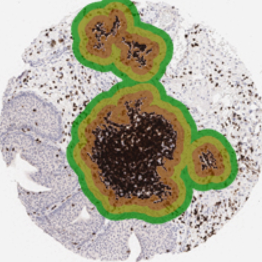
IHC Immune Status in situ APP: Tumor immunology
The IHC Immune Status in situ APP is ideally suited for immunological investigations due to its capability to measure the distance of single cells from tissue metastructures. In this example the APP was applied to bladder cancer Tissue Microarrays.
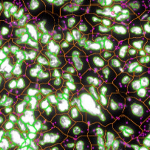
Leishmaniasis APP: Leishmaniasis parasite detection & analysis in cell culture and tissue
The Leishmaniasis APP was designed to assist in immunology research into Leishmaniasis and can be adapted for research on other intracellular parasites. It can be used both on cell cultures and tissue sections.
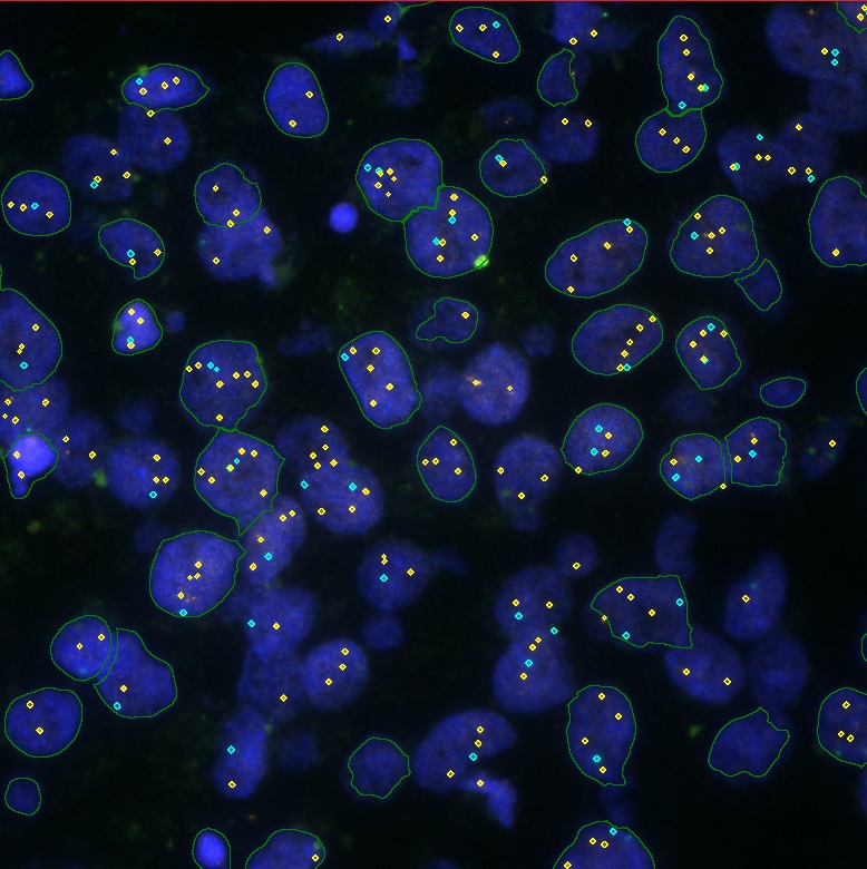
IF Dots APP: FISH
This example shows how the IF Dots APP can be applied to clinical tissue FISH evaluations. The APP can also be applied to any analysis of dot sized objects, in conjunction with all other StrataQuest features. For FISH evaluation the recommended workflow is to run the dot algorithm on all dot stainings and then to select suitable nuclei by their large size and high compactness.


