Brightfield APPs
Brightfield APPs are designed to work on brightfield images and digital slides – usually these will be immunohistochemical stains, but for some requirements (e.g. fibrosis detection) histochemical stains can also be used and quantitatively evaluated. As opposed to immunofluorescence stainings, immunohistochemical stainings need to be color separated before being processed further for image analysis. In the following, find a representative selection of StrataQuest Brightfield APPs.
IHC²
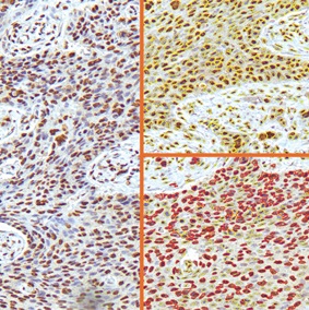
The IHC² APP unmixes two markers (e.g. chromogen and counterstain) in an IHC or HC digital slide and segments single cells into nucleus, and/or perinuclear area and/or cytoplasm. Each segmented cell compartment is measured for up to 20 intensity, statistic and morphometric parameters which are displayed in scattergrams and histograms and can be exported.
APP Category 1
IHC3
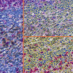
The IHC3 APP unmixes three markers (e.g. two chromogens and counterstain) in an IHC or HC digital slide and segments single cells into nucleus, and/or perinuclear area and/or cytoplasm. Each segmented cell compartment is measured for up to 20 intensity, statistic and morphometric parameters which are displayed in scattergrams and histograms and can be exported.
APP Category 2
IHC Membrane
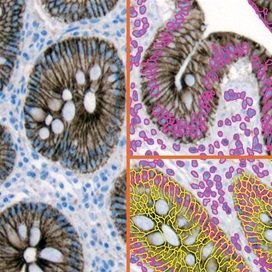
The IHC Membrane APP unmixes up to three markers in an IHC or HC digital slide and segments cells into nucleus, and/or perinuclear area and/or cytoplasm, as well as into membrane (e.g. HER2/neu). Each segmented cell compartment is measured for up to 20 intensity, statistic and morphometric parameters. Three more parameters are measured for membrane intensity and angle of staining. All parameters are displayed in scattergrams and histograms and can be exported.
APP Category 2
IHC Angio
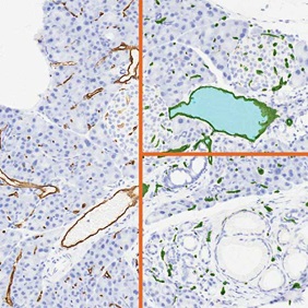
The IHC Angio APP detects blood vessels based on appropriate stains (e.g. CD31) and measures overall vessel area as well as lumen area. The vessel detection also can be set to close open stained vessel walls and to connect separated vessel sections within a definable distance. The APP outputs number and vessel density as well as areas of vessels, endothelium and lumina.
APP Category 2
RNA Scope
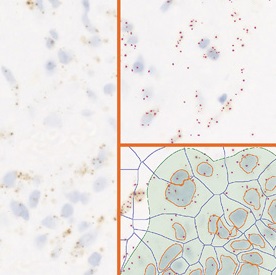
The RNA Scope APP provides dot detection per cell within the nuclear compartment (nucleus and/or cytoplasm) for up to three markers in CISH and SISH experiments. Each segmented cell compartment is measured for up to 20 intensity, statistic and morphometric parameters. Dot parameters are provided per cell and include count, mean intensity, total dot area, and sum of intensity as well as area and intensity lists for all single dots.
APP Category 2
CLASSIFIER
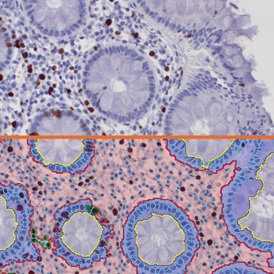
The Classifier APP is based on trainable Deep Learning technology and provides tissue metastructure detection. In the above example it separates the colon tissue into crypts, stroma and infiltration areas. Combined with the IHC2 cellular detection APP the Ki-67+ nuclei in each compartment can be quantitatively analyzed for up to 20 parameters.
APP Category 3
IHC ADIPOCYTE
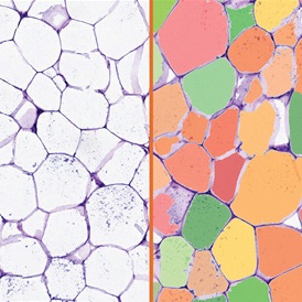
The IHC Adipocyte APP quantifies adipocytes as to their lumen in adequate HE samples. Small rips in adipocyte membranes are mended automatically and cell membrane artefacts in adipocyte lumina are automatically eliminated as are lumina on sample borders. The APP outputs area measurements for all detected adipocyte lumina.
APP Category 1
IHC MACROPHAGES
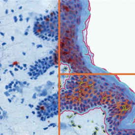
The IHC Macrophages APP detects macrophages based on adequately stained IHC samples. The APP can be combined with area detection and distance range algorithms, in the sample above to determine the distance of Langerhans cells from the border of the epidermis within and without. Each segmented cell compartment is measured for up to 20 parameters, as is the distance of each cell to the boundary.
APP Category 3
IHC MULTI-SHADES
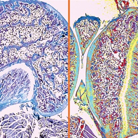
The IHC Multi-Shades APP provides semi-automatic color separation for up to six markers or colors in an IHC or HC digital slide. In the above sample it has been used to detect and segment different levels of ossification based on Azan stain. Each detected area is measured for up to 20 intensity, statistic and morphometric parameters.
APP Category 1
IHC META.CELLS
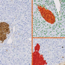
The IHC Meta.Cells APP combines the detection of IHC/HC stained metastructures (e.g. Langerhans islets) with single cell detection (segmentation of cells into nucleus, and/or perinuclear area and/or cytoplasm). Detected cells can be classified and visualized as being either within or outside of detected metastructures. Each detected area and cell compartment is measured for up to 20 intensity, statistic and morphometric parameters.
APP Category 2
IHC IMMUNE STATUS IN SITU
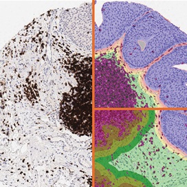
The IHC Immune Status in Situ APP provides phenotypic characterization of immune cells in context with detected metastructures (e.g. tumors, glands). It measures the distance of cellular objects to the metastructure boundary (within and/or outside). Distance ranges can be defined. Each segmented cell compartment is measured for up to 20 parameters, as is the distance of each cell to the boundary.
APP Category 3
PULMO
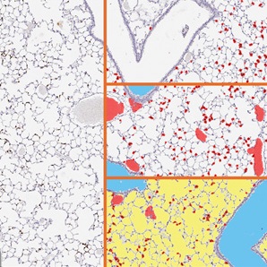
The Pulmo APP segments the metastructure components of lung, including tissue, bronchioles, blood vessels and alveoles. Each segmented metastructure is measured for up to 20 morphometric parameters.
APP Category 3
IHC TUMOR-STROMA
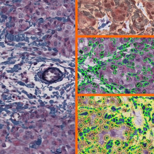
The IHC Tumor-Stroma APP combines the segmentation of tumor and stroma (based on the morphology) and the detection of specifically stained cell populations. It segments the cells into nucleus, and/or perinuclear area and/or cytoplasm. Each segmented cell compartment in tumor and/or stroma is measured for up to 20 intensity, statistic and morphometric parameters which can be displayed in scattergrams and histograms and exported.
APP Category 2
TUMOR FOCI
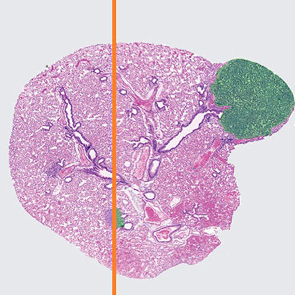
The Tumor Foci APP allows to detect the whole tissue and more important tumor foci based on nuclear structure analysis, mainly on HE staining. The number and area of tumor foci as well as their density is measured.
APP Category 1

