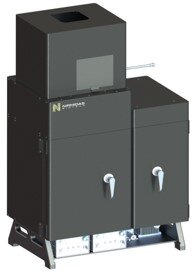
DeepVision™ NIR-II/SWIR
Small Animal Fluorescence Imaging System
In vivo NIR-II/swir Fluorescence Imaging
Fluorescence imaging of small animals using existing commercial instruments detect in the ~800-900 nm range, suffering from shallow imaging depth and high background due to light scattering and tissue autofluorescence. NIR-II/SWIR imaging is a revolutionary development detecting in the 1000-1700 nm range to greatly suppress these effects, affording single cell resolution at ~ 3 mm depth and useful resolving power at up to ~ 1 cm depth. Combined with NIR-II probes (molecules, quantum dots and rare-earth nanoparticles) from Nirmidas Biotech, DeepVision™ can be used for vasculature, tumor, brain, lymphatic/lymph node imaging and molecular imaging, empowering research to interrogate cardiovascular, cancer, brain and immune diseases.

Key Features:
- A high performance and cost effective NIR-II/SWIR fluorescence/luminescence system with multiple, customizable lasers
- The only small imaging system detecting in both NIR-I and NIR-II windows: wavelength of imaging spans 400 nm – 1700 nm with high detection efficiency in NIR-II 1000 – 1700 nm range
- For in vivo small animal imaging, ex vivo and in vitro tissue, organism and cellular imaging
- Low and high magnifications adjustable for whole body and microscopic imaging by switchable lens sets
- Capable of single cell and single micro-vasculature imaging resolution in vivo
- Up to 120 frames per second video rate imaging
- Lifetime imaging capability

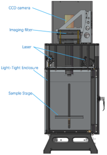
Inside the DeepVision™:
CCD Camera
- The DeepVision™ CCD camera is ~10 mm x 8 mm with 640 x 512 pixels for high imaging resolution
- 400 – 1700 nm with high detection efficiency
- CCD TE and water cooling to ensure low dark current and low noise
Imaging Chamber
- Light-tight imaging chamber
- 808 nm and 980 nm lasers (customizable)
- Ex/Em filter wheels – 4 filters each
- Heating pad for mouse
- Adjustable imaging field, for whole body or microscopic imaging of mice
- X-Y-Z mouse stage with automated control
Use of DeepVision™ NIR-II Small Animal Fluorescence Imaging System
Through scalp/skull non-invasive video rate imaging of cerebral vessels in a mouse head. The fluorescence signal of rare earth nanoparticles in the cerebral vessels was observed through the scalp and skull of mice. During the video recording, the mouse scalp was slightly moved, and the transparency and three-dimensional structure of the mouse head were clearly visible.


