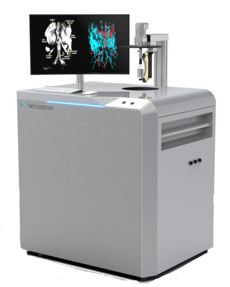
Heliosonix

Heliosonix AREA
Heliosonix AREA

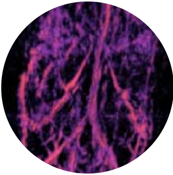
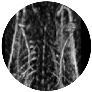
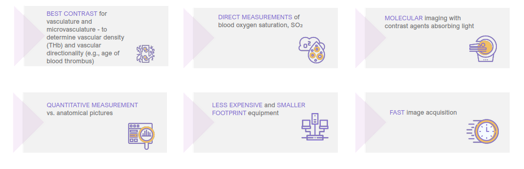
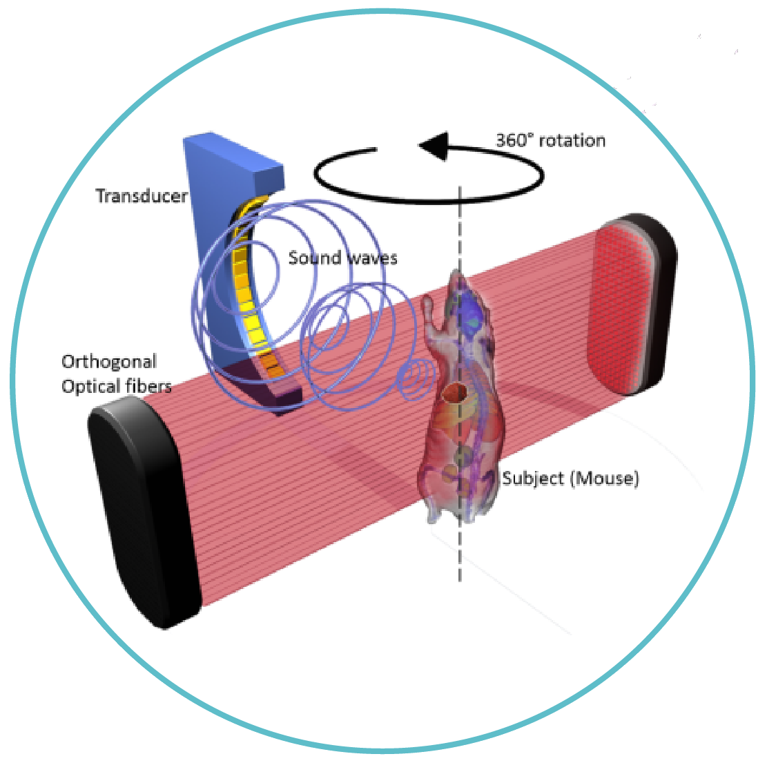
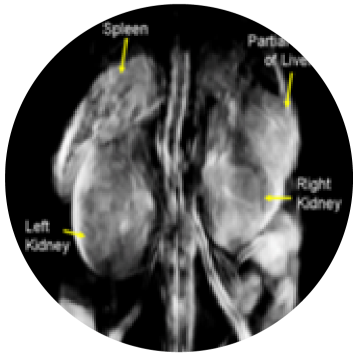
When a short-pulsed laser energy penetrates the tissue, it heats and expands, generating thermo-acoustic (ultrasound) waves, which can be reconstructed to show the distribution of optical absorption inside the tissue.
Keeps mice alive – under anaesthesia
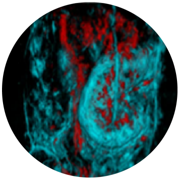
Obtains live 3D images with high resolution of inner structures,
including organ, vasculature and skeleton systems
Captures bones, soft tissue organs and blood flow
Delivers results within in 3-4 min
Allows to quantitatively track the effect of pharma
treatment over time in the same animal

AREA does not require regulatory approval
Buyers: universities, CROs, biopharma
Numerous prototype delivered and installed
Commercial sales launched in 2020 and 2 systems
sold and installed in China
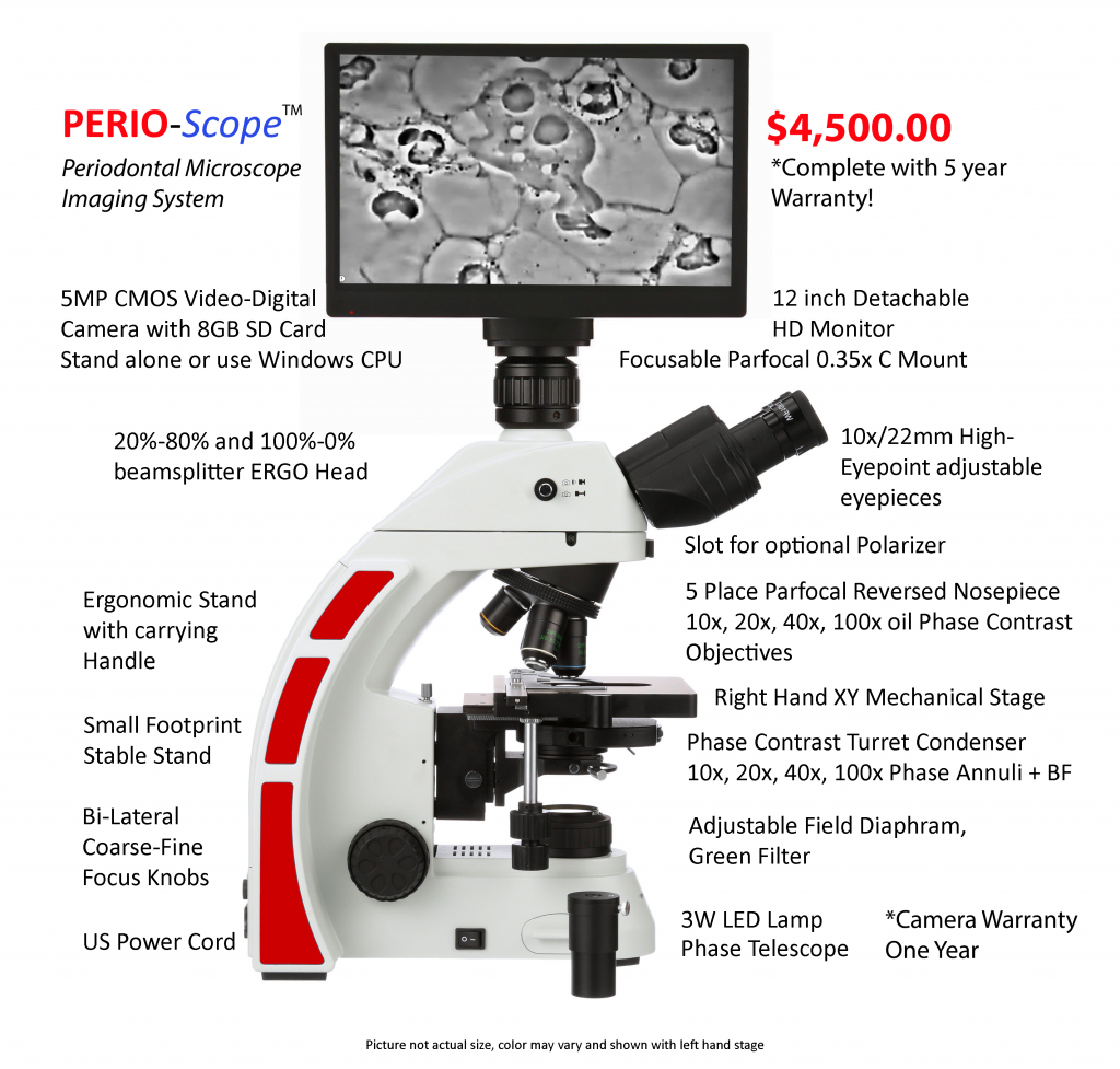
Announcing PERIO-Scope

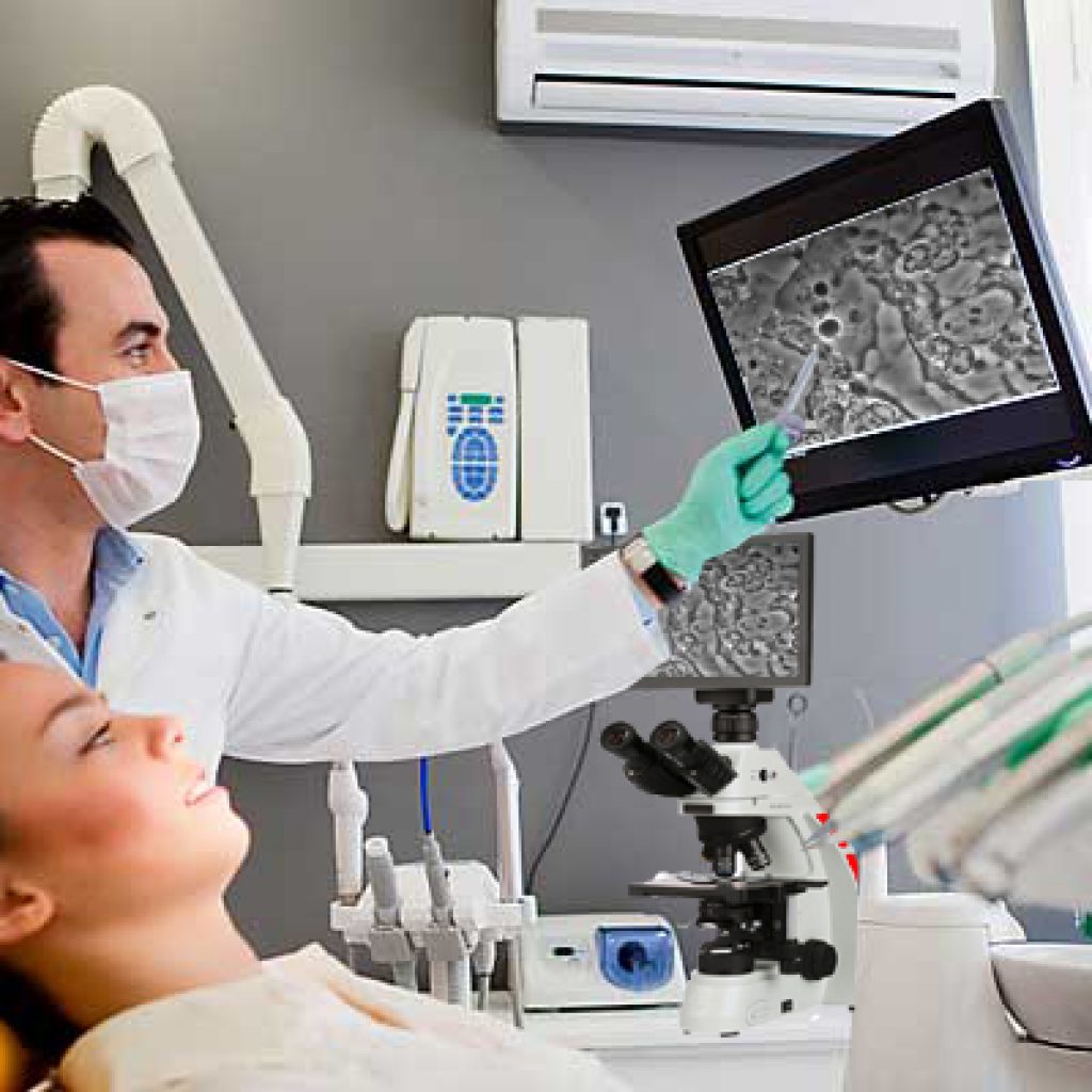
PERIO-Scope microscope imaging system for PERIODONTISTS

Our new product is designed specifically for periodontists to help prevent further gum disease by showing their patients how to achieve a healthy mouth with ‘LIVE’ video imaging. “If a picture is worth a thousand words, video is worth a million, seeing is believing”

  • New way to monitor and help eliminate periodontal gum disease.
  • Easy to use and amazing to watch your patient’s reaction!

The PERIOScope is designed to allow patients to see these living organisms in their own mouths.

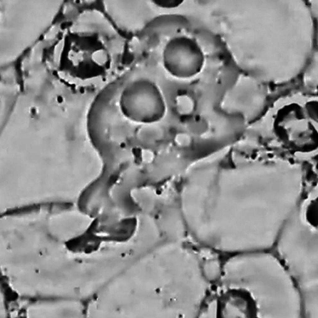
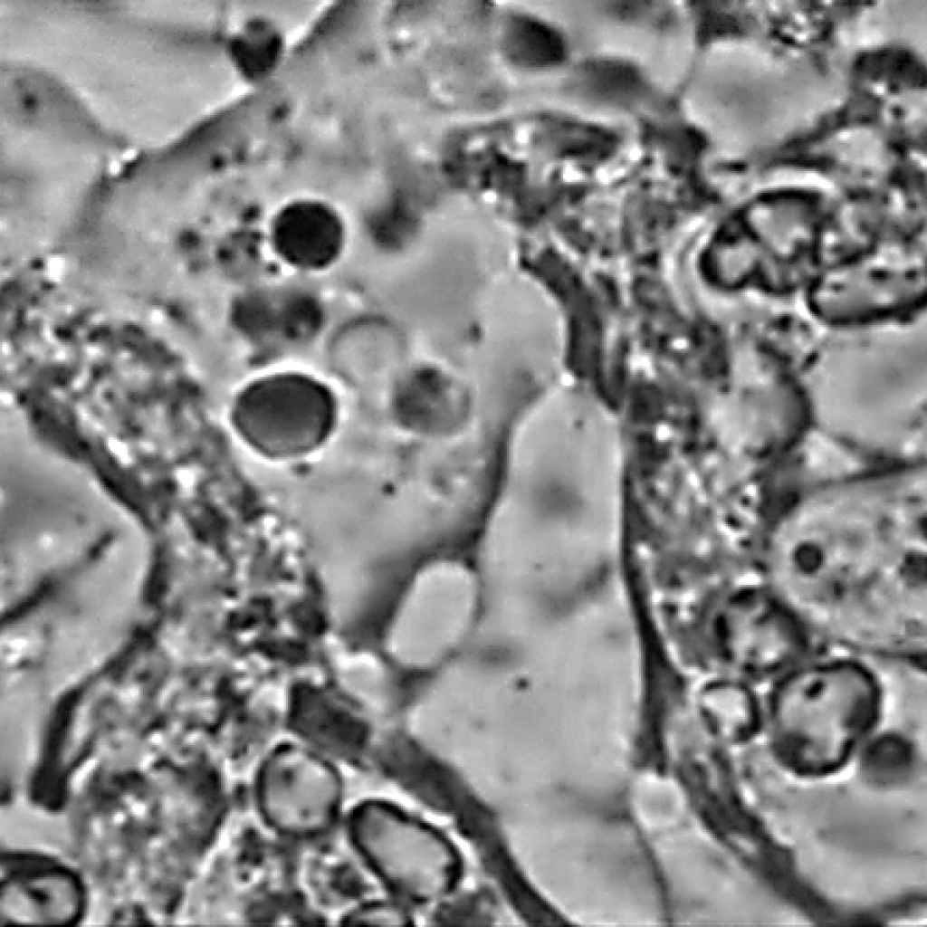
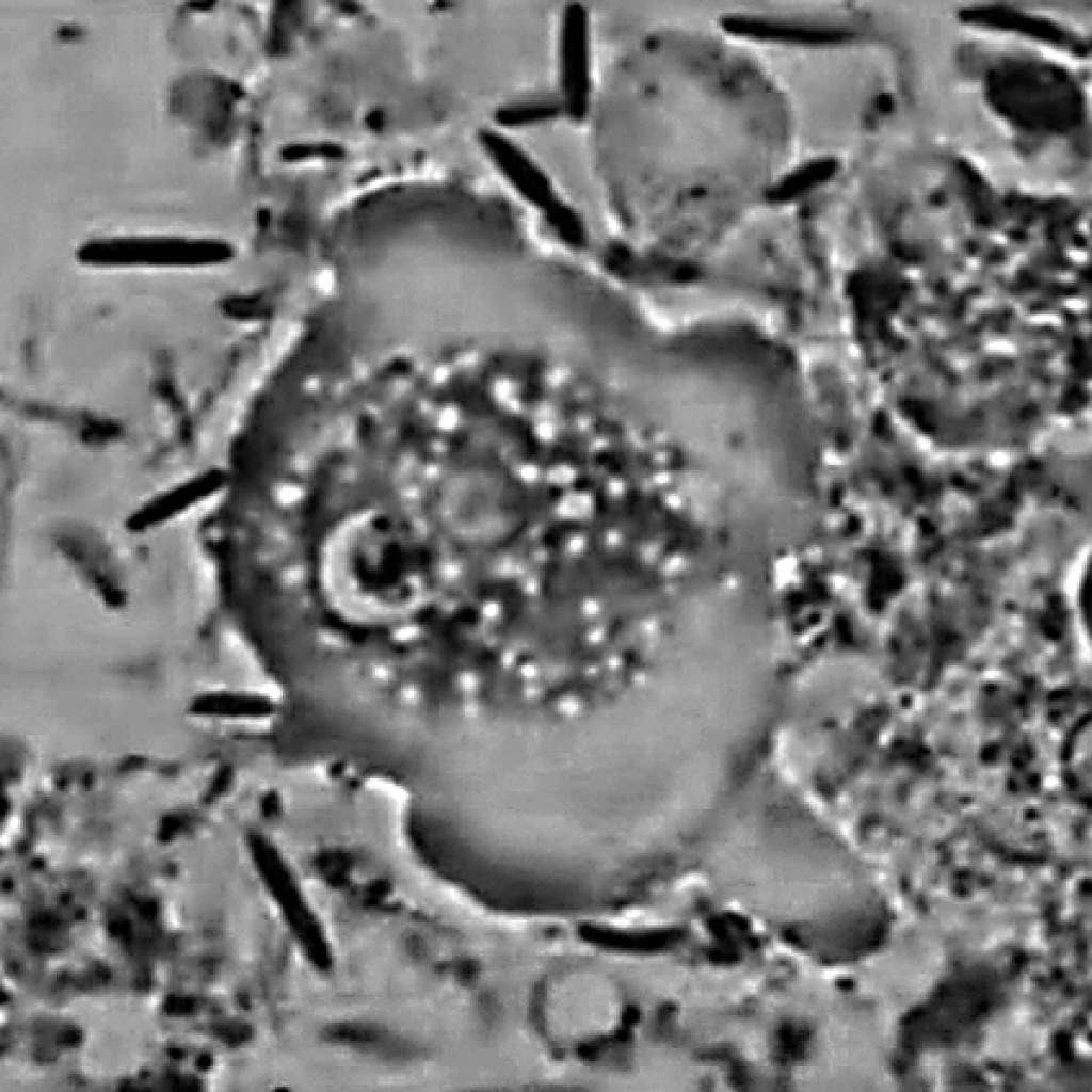
Periodontal Disease treatment can be visually explained to your patients with the help of a dedicated microscope and video camera capable of recording live video and still image capture. When images of epithelial cells, bacteria, red and white blood cells, trichomonas and even amoebas are shown to patients they can see for themselves the severity of gum disease.

Our PERIOScope phase contrast microscope imaging system is a simple, effective way for periodontists to show the ill effects of gum disease. Using “LIVE’ video feed, patients can experience for themselves the long-term effects of poor dental hygiene.

Using our specialized imaging tools, periodontists can visualize, record and track the progress of treatment. Operation is easy and fast. When used in conjunction with large HD monitors the images almost jump off the screen for patients and health providers benefit and understanding.

The PERIOScope comes complete and ready to use stand-alone with the included 12-inch HD monitor or with a Windows or Mac computer connected via USB2 cable. Beautiful live, phase contrast images of unstained saliva showcase all manner of material found in the patient’s mouth and gums.

Motic Launching FS-Live

Call Meyer Instruments for the Motic FS Live

Motic Launching FS-Live Telepathology System for Frozen Sections

Motic announces the FS-Live Telepathology System for frozen cases or fine-needle aspiration. This advanced, microscope-based whole slide imaging system provides high quality live images efficiently to pathologists from anywhere in the world, specifically for remote diagnosis and interpretation. The FS-Live eliminates the need to send pathologists and other health specialists to remote hospitals solely to read these cases. The system allows any pathologist on the network to view a case slide in a matter of seconds. Available with single or 6 slide capacity.

“Motic is relentless in perfecting the art of telepathology and digital pathology,” said Alex Lobozar, Motic General Manager, Sales and Marketing for North America. “We are constantly exploring ways to improve our instruments to be increasingly user-friendly and accessible for all.”

Key Features

Continuous Autofocus – unique to ALL scanners on the market, SUPER time saver

Live Mode 2x, 4x, 10x, 20x, 30x, 40x – duplicates common magnifications

Moisture Resistant – can be used with wet slides

Simple Operation – no complicated instructions to learn

Keyboard, Mouse or Hot Key Controls – multiple ways to focus or change magnifications

Heat Map – keep track of where you have viewed and at what magnification

Acts as a traditional scanner as well – save slides in multiple formats SVS, TIFF, JPEG, JPEG 2000, MDS


HREM – New 3D Imaging

HREM 3D

Call Meyer Instruments at 281-463-0342 for more information about this amazing 3D Digital Imaging System.

High Resolution Episcopic Microscope (HREM)

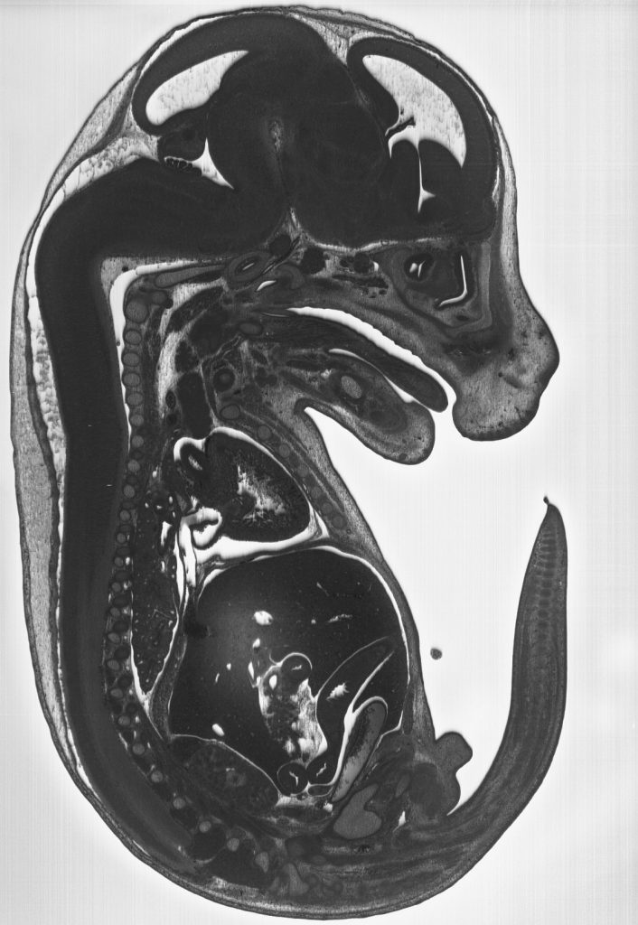
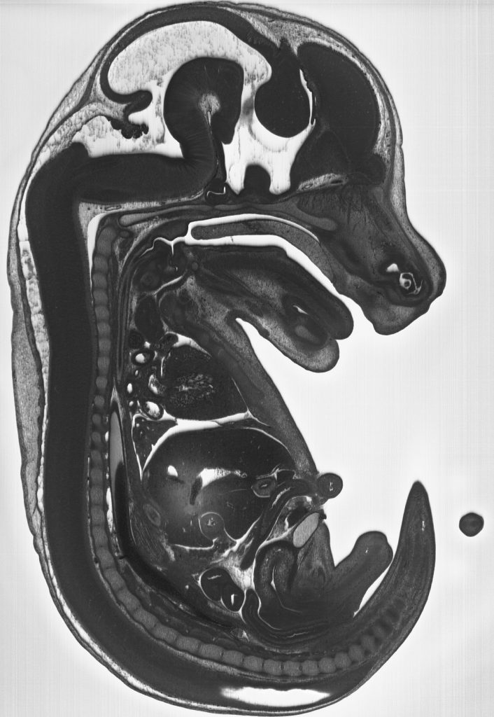
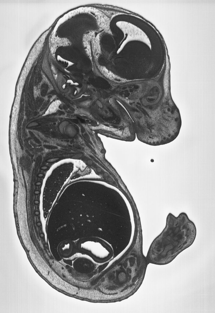
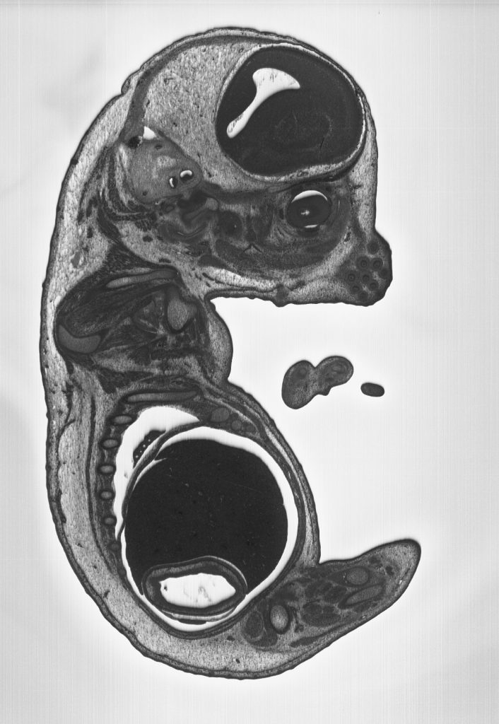
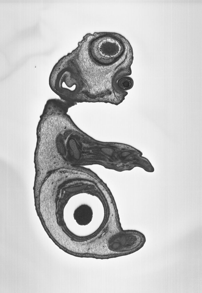
The optical High Resolution Episcopic Microscope (HREM) is a 3 dimensional image generation device for whole histological samples or embryo’s.

Consisting of a motorized microtome, fluorescence stereomicroscope and camera, the HREM cuts away a sample at 1-5 micron intervals and photographs the remaining faceplate, section by section. The result is hundreds or thousands or perfectly aligned images that are available for reassembling in 3D. Maximum sample size is 25mm in diameter (although they are experimenting with a new 40mm holder as well)