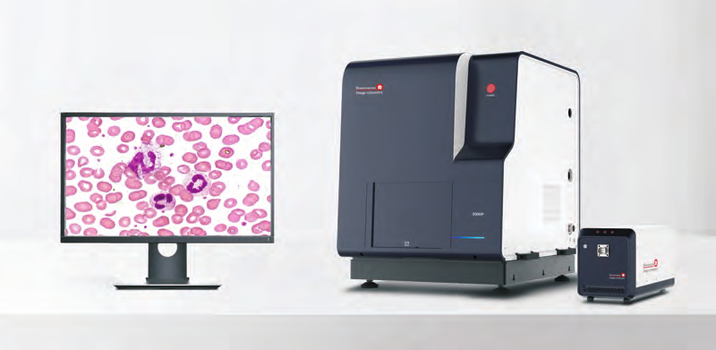
Ultra-High Speed Scanning
for Cytology and Hematology
* Unprecedented, Ultra-High speed oil scanning (under 3 minutes for 20mm x 40mm area)
* Scan whole slides and selected regions of interest with air and oil objectives up to 100x.
* Automatic oil dropping and retrieving
* 5 place manual or motorized nosepiece
* Accommodates two 1 x 3 inch glass slides
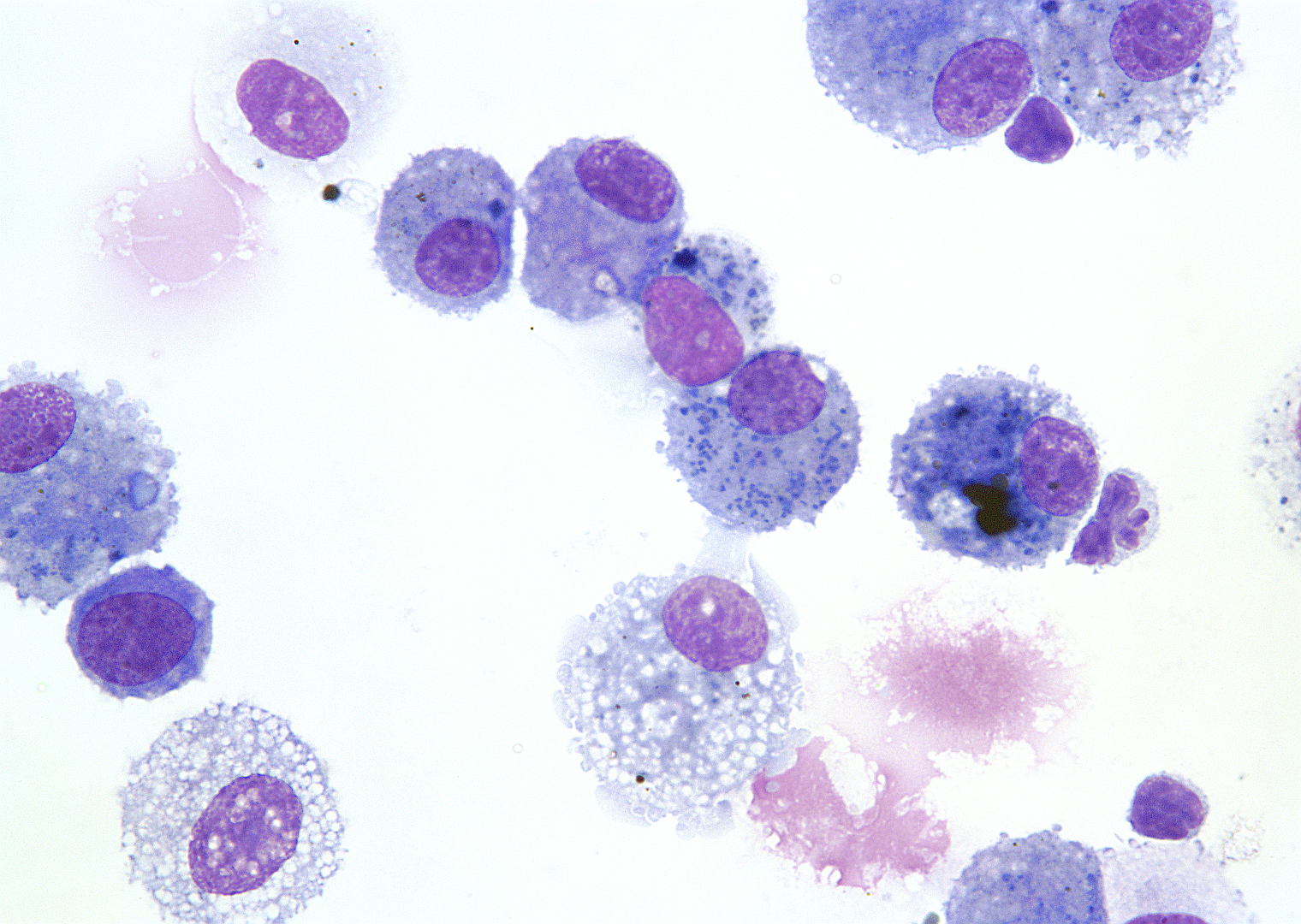
Slide Scanning times for
dry & oil objectives
(20mm x 40mm area)
*Less than 1 minute for 40x at 0.25 um resolution
*Less than 2 minutes for 60x at 0.17 um resolution
*Less than 3 minutes for 100x oil at 0.1 um resolution
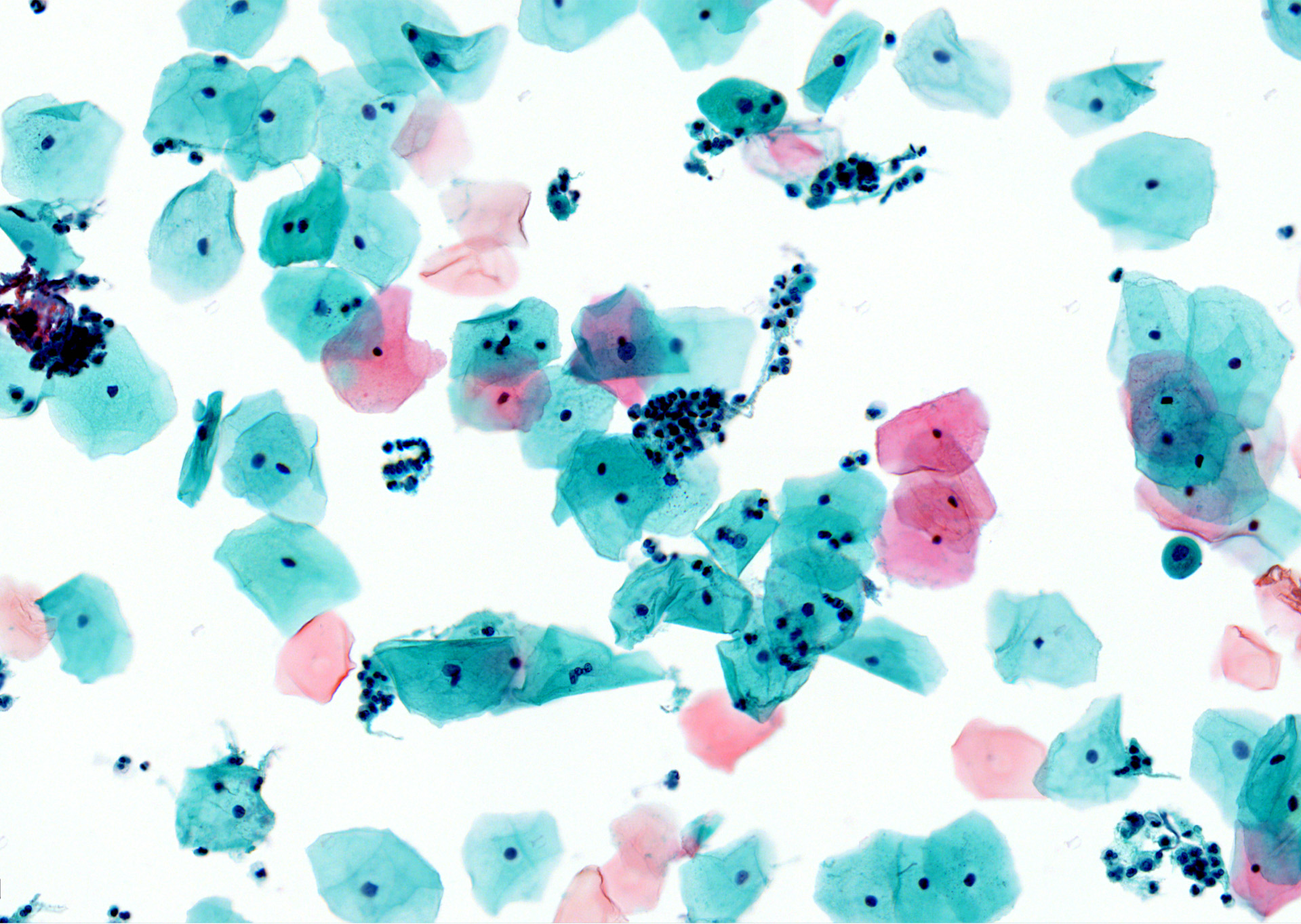
Image Browsing
*Remote control from anywhere at anytime and simultaneously work with colleagues for second opinions.
*JEPG or SVS image format, 0.1um resolution picture, file size up to 10 gigabytes *Seamless picture stitching, zooming in or out from 0.25x-100x
*Annotation or measurements on regions of interest
*When scanning at 0.1um resolution, the whole slide is composed of 14,000 pictures with 5 million pixels of each
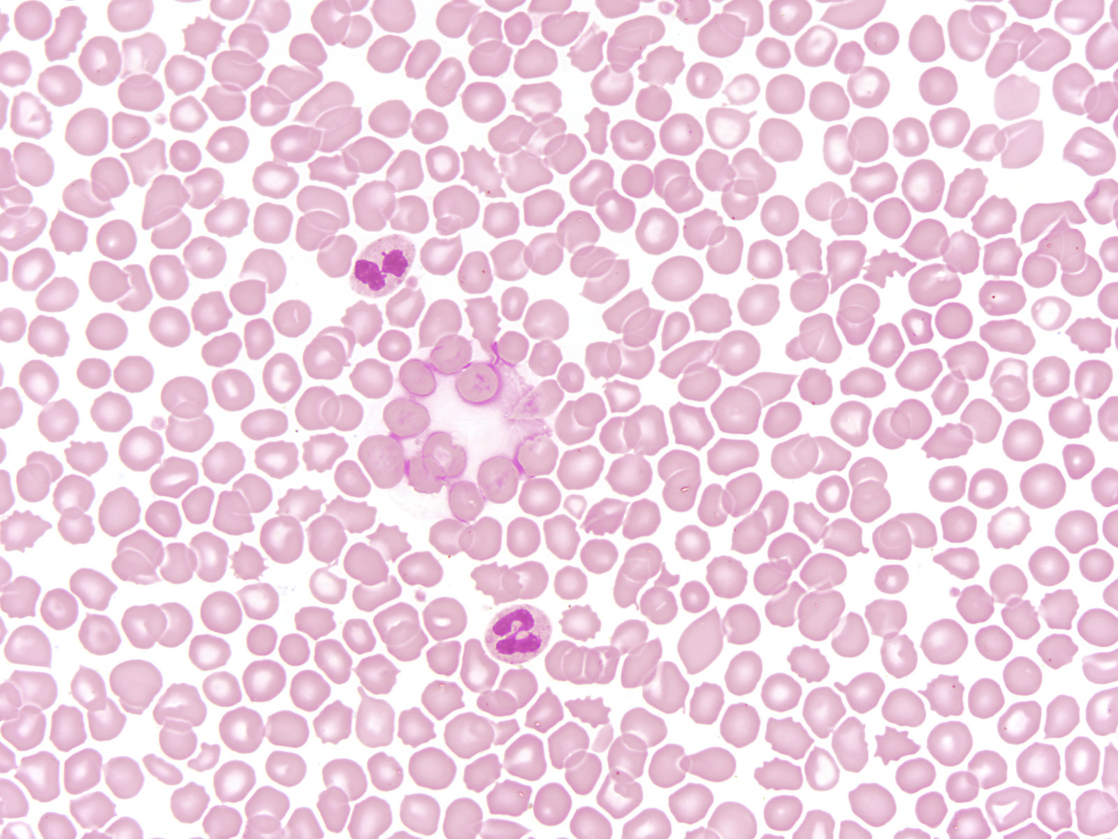
Deep Learning
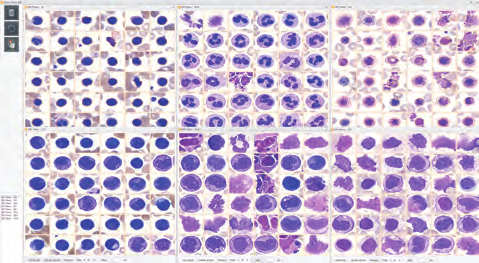
*Optional software for image quantification available for various morphological indexes
*Nuclear area, nuclear perimeter, nuclear circularity, staining brightness of nuclei, nucleus cytoplasm ratio,
*Cytoplasmic area, nucleus cytoplasm ratio, cytoplasmic granularity, cell circularity, cytoplasm staining brightness and similarity coefficient of deep learning classification
*Neural net allows deep learning practices
*Up to 4 GPU’s are utilizes in high performance workstations
*Categorize 4,000 frames per second with 64×64 pixels areas (cells or other objects)
Data Viewing
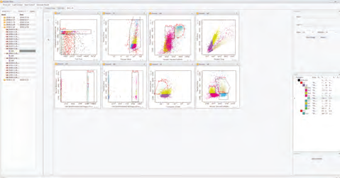
*Data in formats of scatter diagram, histogram, high dimensional graph.
*After 14,000 pictures with 5M pixels are converted to quantitative data of the sample, multiple scatter diagrams or histograms will be displayed within several seconds for observation and study
*Data is saved in FACS3.0 format, can be opened using FACS analytical software, and data file of 10G can be converted into several mega FACS data file.
Application Areas
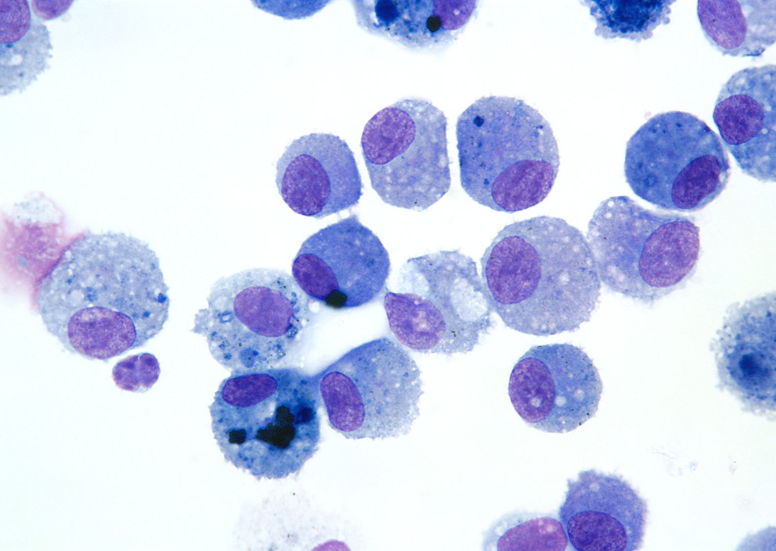
*Cytology
*Hematology
*Microbiology
*Pathology
*Oncology
*Cell Biology
*Veterinary Medicine
Bionovation Models |
CSFA350-40 |
CSFA350-60 |
CSFA350-Plus |
CSFA800-Basic |
| Camera-Megapixel | 5MP | 5MP | 5MP | 5MP |
| Camera-Frames per second | 75 fps | 75 fps | 75 fps | 163 fps |
| Motorized Nosepiece | no | no | no | yes |
| Motorized LED Illuminator | no | no | no | yes |
| Auto oil drop | no | no | no | yes |
| Objective-100x oil | no | no | 100X oil n.a. 1.25 | 100X oil n.a. 1.25 |
| Objective-60x | no | 60X n.a. 0.8 | 60X n.a. 0.8 | 60X n.a. 0.8 |
| Objective-40x | 40X n.a. 0.65 | 40X n.a. 0.65 | 40X n.a. 0.65 | 40X n.a. 0.65 |
| Adaptor | 0.35x | 0.35x | 0.35x | 0.35x |
| Microns per pixel – 100x | no | no | 0.10 | 0.10 |
| Microns per pixel – 60x | 0.17 | 0.17 | 0.17 | 0.17 |
| Microns per pixel – 40x | 0.25 | 0.25 | 0.25 | 0.25 |
| Slide capability | 2 | 2 | 2 | 2 |
| Scanning time-100x | no | no | <6 min for 20mm x 40mm | <3 min for 20mm x 40mm |
| Scanning time-60x | no | <4 min for 20mm x 40mm | <4 min for 20mm x 40mm | <2 min for 20mm x 40mm |
| Scanning time-40x | <30 sec for 15mm x 15mm | <30 sec for 15mm x 15mm | <20 secs for 15mm x 15mm | <20 secs for 15mm x 15mm |
| Computer – GPU | 1 GTX1660TI GPU | 1 GTX1660TI GPU | 2 GTX1660TI GPU | 2 RTX2060s GPU |
| Computer – CPU | AMD/0.24T | AMD/0.24T | AMD/0.24T | Intel/0.5T |
| Computer – Hard Disk Drive | 1T | 1T | 1T | 6T |
| Remote control | Yes, via Team Viewer | Yes, via Team Viewer | Yes, via Team Viewer | Yes, via Team Viewer |
| AI – tissue scanning | Yes | Yes | Yes | Yes |
| AI – Peripheral blood smear | Yes | Yes | Yes | Yes |
| AI – cytology | No | No | No | Yes |
| AI – bone marrow | No | No | No | Yes, 15 mm x 15 mm |
| AI – cells analyzed per second | No | 100 | 200 | 800 |
| Regulatory (pending) | CE, UL | CE, UL | CE, UL | CE, UL |
FINALLY AN ULTRA-FAST SLIDE SCANNER FOR CYTOLOGISTS AND HEMATOLOGISTS!
Please contact us for a WebEx demonstation. We also offer scanning
services for your slides at any resolution (0.1um~0.45um) or
customized deep learning programs for your study.

